
My Alzheimer's Journey
Part 29 - Everything There Is To Know About An MRI

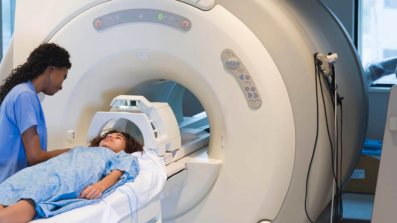
I’m scheduled for an MRI Tuesday, November 4. This will be the first of many MRIs for me while I am receiving Lecanemab. The purpose of these MRIs is to check for brain swelling (ARIA-E) and brain bleeding (ARIA-H). I really don’t like getting an MRI. The noise and being restrained drives me nuts.
I decided to learn more about the MRI machine and the scanning process in the hope that it will put me more at ease. Of course, I am sharing my findings with you.
MRI stands for Magnetic Resonance Imaging. An MRI is imaging technology that produces three-dimensional, detailed anatomical images. It is used for disease detection, diagnosis, and treatment monitoring.
The National Institute of Biomedical Imaging and Bioengineering provided a very comprehensive description of how an MRI works. I copied and pasted their description below. The bold text is my attempt to use plain English.
“Using powerful magnets, the MRI machine produces a strong magnetic field that forces protons (the human body is made mostly of water, and each hydrogen atom in water has a single proton) in the body to align with that field (the protons are told to line up with the magnetic field). When a radio frequency current is then pulsed through the patient, the protons are stimulated (they absorb energy), and spin out of equilibrium, straining against the pull of the magnetic field (the protons jump out of line and turn ninety degrees from the magnetic field). When the radio frequency field is turned off, the MRI sensors are able to detect the energy released (protons shed their energy) as the protons realign (get back in line) with the magnetic field.”
The released energy (radio signals) contains information about the protons’ position in the body, as well as their environment (such as healthy tissue or tumors). A computer processes these varying signals and translates them into a detailed, cross-sectional image that distinguishes between different tissues.
In order to create an image, the MRI machine has to repeat the process of firing radio frequency pulses many times. This is what makes the jackhammering type noise. Each type of scan (or pulse) has its own special sound. The MRI machine uses different sequences like 2D and 3D T1W, T2W, Diffusion, and more, along with various orientations such as axial, coronal, oblique or sagittal. These different sequences create images with different contrasts, angles, and coverage areas.
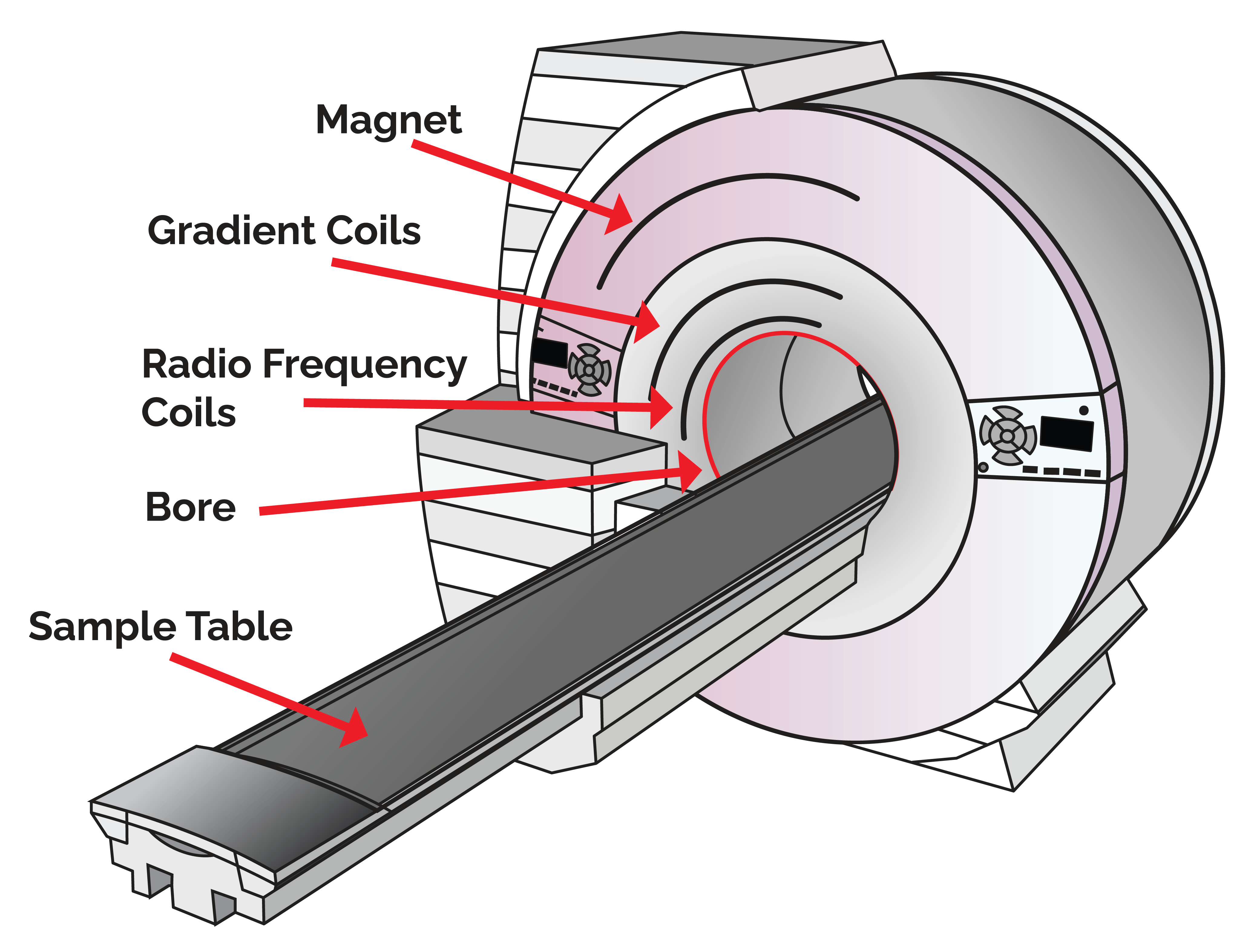
The magnet is extremely cold and surrounded by liquid helium. Magnetic field strength is measured in gauss units. The Earths magnetic field is .5 gauss. MRI machines use fields between 3000 and 70,000 gauss. This is why you can’t have metal in an MRI room. A very tragic and sad event happened in July 2025 in New York when a woman called her husband to come into the room after her scan. He was wearing a heavy metal chain necklace. He was sucked into the MRI machine. Instant death.
Technicians always provide ear plugs and headphones to protect your ears during an MRI. Trust me, it is still very loud.
Siemens (a leading manufacturer of MRI machines) has developed advanced noise reduction technologies. This noise reduction technology is present on all MAGNETOM scanners. The price of a new MAGNETOM machine starts at $1.6 million and goes up to $2.2 million. Plus another $500,000 for installation. Ouch!
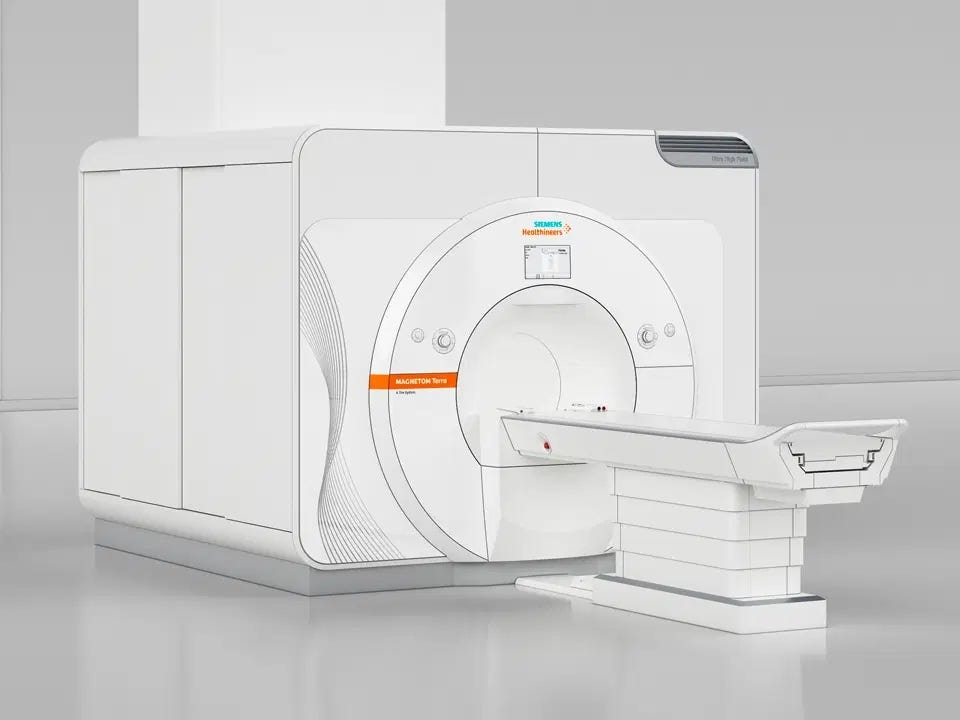
I checked with Duke and I will get my MRIs in a 3T Siemens Verio machine. Some models of this machine include noise reduction technology. I hope my machine has it!